


Osteoarthritis is a chronic pathology that affects the connective tissue structures of the musculoskeletal apparatus. For the disease characterized by progressive course with the gradual destruction of cartilage. Arthrosis is diagnosed in most patients after 65 years as one of the reasons for its development is the natural aging process.
To the appearance of degenerative-dystrophic pathology of the result of preceding trauma, endocrine and inflammatory diseases, excessive physical exertion or, on the contrary, a sedentary lifestyle. Leading symptoms of osteoarthritis are pain in the joint swelling, limitation of movements.
To diagnose pathology conducts instrumental studies — radiography, arthroscopy, MRI, CT. Arthritis 1 and 2 severity treated conservatively course taking drugs, physical therapy and massage, exercise therapy. When irreversible destructive changes in the joints, surgical intervention — arthrodesis, arthroplasty.
Pathogenetic mechanisms
In osteoarthritis occur pronounced changes in the internal connective tissue structures. On cartilaginous tissue formed deforming erosion, which causes destruction of the collagen fibers, and proteoglycans consisting of a protein (5-10%) and glycosaminoglycans (90-95%). The result of collagen mesh loses its stability, begin to be released metalloproteinases that destroy all types of extracellular matrix proteins. Degradation is accelerated by increasing the biosynthesis of collagens and stromelysin. Usually normal values of enzymes controlling cytokines — small peptide information molecules. But with the progression of osteoarthritis, the concentration of these proteins decreases, which causes the release of a large number of enzymes damaging cartilage.
Proteoglycans with altered structure begins to absorb water molecules, which are unable to keep. Therefore, the excess fluid enters the collagen fibers. They "swell", lose strength and elasticity. Qualitative and quantitative composition of synovial fluid is also experiencing negative changes. In osteoarthritis it reduces the concentration of hyaluronic acid. To the hyaline cartilage cease to be sufficient for their regeneration the amount of nutrients and oxygen. In the cartilaginous tissues formed foci of softening, and then cracks, specific necrotic growths. Bone head bare, you begin to microtraumatic at offset relative to each other.
Causes and predisposing factors
The causes of primary (idiopathic) osteoarthritis is not yet established. It occurs when the absence of any precipitating factors, therefore, put forward the theory of hereditary predisposition to premature breakdown of cartilage. Secondary osteoarthritis develops as a consequence of other pathologies of joints or previous injuries. What can cause degenerative diseases:
- injury to the joint or nearby connective tissue structures — fracture, dislocation, injuries to the meniscus, a partial tear of muscles, ligaments, tendons, or their complete detachment from bone base;
- congenital dysplastic developmental disorder of articulation;
- violation of the endocrine glands, disorder of metabolic processes;
- rheumatism, or rheumatic fever;
- rheumatoid, reactive, metabolic, psoriatic, or gouty arthritis, polyarthritis;
- purulent arthritis provoked by streptococci, or Staphylococcus epidermidis staphylococci;
- tuberculosis of any localization, brucellosis, chlamydia, gonorrhea, syphilis;
- degenerative disease, for example, dissecting Legg.
To the development of osteoarthritis predisposes hypermobility of the joints due to the production of a particular collagen. This condition is diagnosed in 10% of the planet's inhabitants and is not considered a pathology. But the hypermobility is accompanied by weakness of the tendinous-ligamentous apparatus, which leads to frequent injuries, special ankle (sprains and torn ligaments, sprains).
Cause arthritis sometimes are disorders of hematopoiesis, for example, hemophilia. Hemarthrosis, or bleeding into the cavity of the joint, provoking the deterioration of the trophic cartilage and their destruction.
By the predisposing factors include old age, frequent stress on the joints that exceed the limits of their strength, weight gain, surgery, and hypothermia.
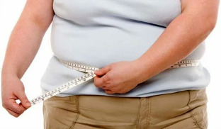
The risk group includes women in menopause, people living in adverse environmental conditions or contact with toxic chemical compounds. Deficiency in the diet of foods with vitamins and minerals, creating conditions for the gradual degradation of the hyaline cartilage.
The clinical picture
The danger of osteoarthritis is the lack of symptoms in the first stage of its development. Pathology is clinically manifested gradually, the first signs arise against significant destruction of the cartilage tissues. First, the person feels mild pain, without clear localization. It appears after physical exertion — lifting weights, sports training. Sometimes the first clinical manifestation is a crunch, clicks when bending or unbending of the joint. People began to notice that certain movements are difficult. However, at the initial stage of osteoarthritis stiffness occurs in the morning hours and soon disappears.
The disease progresses pain is felt at night, causing not only a sleep disorder, but the emergence of chronic fatigue. The intensity of pain syndrome in the second stage increases with the change of weather, exacerbation of chronic diseases, infection. Markedly reduced range of motion. The reason for the stiffness becomes thinning of cartilage and deliberate restriction of a person's movements in an attempt to avoid pain. This leads to increased load on the opposite joint, which causes further damage. For osteoarthritis characteristic and other specific symptoms:
- pain provokes spasms of skeletal muscles and development of muscle contractures (restriction of passive movements of the joint);
- the crunch in joints, clicks, crackle on the move become permanent, occur on nearly every displacement of the bones relative to each other;
- often, there are painful muscle cramps;
- the joints are deformed, which leads to the disorders of posture and gait;
- in the third stage of arthrosis deformation is expressed so vividly that the joint is bent, and the volume of movements in them significantly reduced or completely absent;
- in the third degree osteoarthritis of the knee, ankle, hip joint when moving the patient uses a cane or crutches.
If untreated, the pathology progresses, and in its course of remission alternate with recurrences, and the frequency of exacerbations increases all the time. Stiffness in the morning, now does not disappear for a long time, it becomes permanent.
Examining a patient with osteoarthritis of 1 degree, the doctor noted only a slight swelling of the joint and complete safety of the range of motion. In the pathology of 2 degrees palpation reveals pain and weakly expressed deformation. In the region of the joint space observed the formation of bone nodules.
For osteoarthritis characterized by the development of synovitis — inflammation in the synovial membranes of the hip, knee, ankle, shoulder joints. Their leading symptom becomes rounded education seal in the field joint, pressing on which there is a displacement of fluid (fluctuation). For acute synovitis may be accompanied by a rise in temperature to 37-38°C, headaches, digestive disorders.
Diagnosis
The diagnosis put on the basis of results of instrumental investigations, clinical features, anamnesis, complaints of patients. General analysis of blood and urine uninformative — all values remain within normal limits if the arthritis is not triggered by metabolic disorders. With the development of synovitis increases the erythrocyte sedimentation rate (30 mm/hour) in the blood increases the level of leukocytes, and fibrinogen. This indicates that occur in the body acute or chronic inflammatory process. Changes in biochemical and immunological parameters occur in secondary forms of osteoarthritis.
The most informative method of diagnostics of degenerative-dystrophic pathology — radiography in frontal and lateral projections.
| Stage of osteoarthritis in accordance with the classification of Kellgren-Lawrence (1957) | Radiographic signs of pathology |
| Initial | The absence of radiological signs |
| First | Indistinct, irregular narrowing of the joint space. A small flattening of the edges of the bone plates, the initial formation of osteophytes or lack thereof |
| Second | Marked narrowing of the joint space greater than the norm by 2-3 times, the formation of a large number of osteophytes, subchondral osteosclerosis. The appearance kistevidnyj enlightenment in the epiphyses |
| Third | The appearance of expressed subchondral osteosclerosis and large marginal osteophytes, significant narrowing of the joint space |
| Fourth | The formation of coarse massive osteophytes, the almost complete fusion of the joint space, the deformation and compaction of the epiphyses of the bones forming the joint |
If after studying x-ray images to the doctor to have doubts in setting the diagnosis, prescribe a CT scan. And to assess the state located around the joint connective tissue structures MRI. When using contrast in dynamics to evaluate blood flow to the tissue, set the stage of the inflammatory process in the development of synovitis.
The main methods of therapy
Osteoarthritis is still incurable disease, as there are no pharmaceutical drugs for the regeneration of cartilage. The main task of treatment becomes the prevention of progression of diseases, preservation of joint mobility. Treatment is long and complex, using both local and systemic medications. Patients should avoid serious stress on the joint, if necessary, to limit the range of motion orthopedic devices such as braces, elastic bandages. Patients with excess weight, you need to make adjustments to the diet for a gradual weight loss and diet.
After achieving a sustained remission patients shown daily physical therapy sessions. The first training is conducted under the guidance of a physical therapist, then the patient performs exercises at home. Physical therapy can be supplemented with swimming, yoga, Cycling.
To reduce the severity of pain prescribers various clinical and pharmacological groups:
- nonsteroidal anti-inflammatory drugs in the form of ointments, tablets, solutions for parenteral administration the active ingredients;
- injections into the joint solutions of anesthetics in combination with corticosteroids;
- muscle relaxants to eliminate muscle spasms and restrictive contractures.
In the therapeutic scheme includes the B vitamins, sedatives, if necessary, tranquilizers and antidepressants. Is also appointed chondroprotectors for long course receiving. This is the only group of drugs that have the ability to partially restore cartilage.
To enhance their clinical activity carried physiotherapy — laser therapy, magnetic field, UHF-therapy.
Any joint pain should be a signal for immediate treatment to the doctor. Therapy conducted at the initial stage of osteoarthritis, it will help stop the destruction of cartilage, to avoid the loss of health and disability.